Vaginal leiomyoma: a rare vaginal tumor
Vol. 66 No. 4, 2025
ROMANIAN JOURNAL of MORPHOLOGY and EMBRYOLOGY
Ciprian-Andrei Coroleuca, Bogdan-Catalin Coroleuca, Alexandra Irma Gabriela Bausic, Diana Elena Soare, Andrei Manu, Cristina Maria Iacob, Mihaela Arina Banu, Maria Victoria Olinca, Aniela-Roxana Noditi, Antoine Edu, Elvira Bratila, Octavian Munteanu
Background/Objectives: While uterine leiomyomas are one of the most common pathologies encountered in gynecology, myomas arising from the vagina are rarely identified and a limited number of cases have been reported so far. With variable locations, diagnosis and surgical treatment of the vaginal leiomyoma can be challenging. Case presentations: We present two cases of vaginal myomas. The first case was of a 43-year-old patient who presented dyspareunia. Clinical examination revealed a vaginal mass, of 2/1 cm, located on the posterolateral left vaginal wall. Ultrasound showed a well confined, hyperechogenic round mass. After vaginal excision and histopathological analysis, the final diagnosis was established - vaginal leiomyoma. The second patient presented it for a routine gynecological examination. A firm nodule was palpated in the posterior vaginal fornix. After transvaginal excision, the diagnosis was histopathologically confirmed - vaginal myoma. Conclusions: Whenever confronted with a vaginal mass, the diagnosis of a vaginal myoma should be kept in mind, as transvaginal excision is the preferred surgical treatment.
Corresponding author: Andrei Manu, University Assistant, MD, PhD Student; e-mail: andrei.manu16@yahoo.co.uk
DOI: 10.47162/RJME.66.4.01 Download PDF Vaginal leiomyoma: a rare vaginal tumor PDF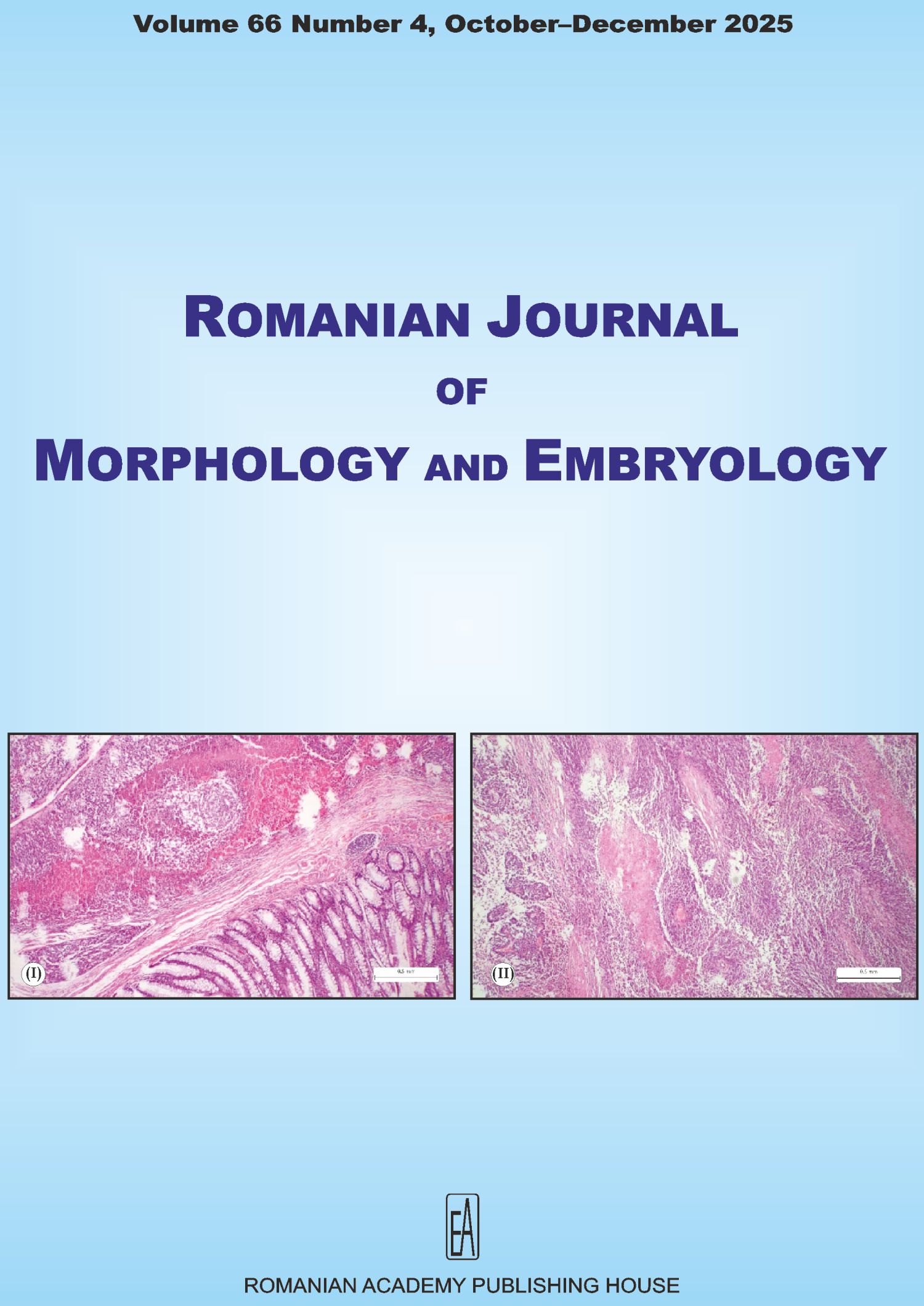 Download cover
Download contents
Journal archive
- vol. 66 no. 3, 2025
- vol. 66 no. 2, 2025
- vol. 66 no. 1, 2025
- vol. 65 no. 4, 2024
- vol. 65 no. 3, 2024
- vol. 65 no. 2, 2024
- vol. 65 no. 1, 2024
- vol. 64 no. 4, 2023
- vol. 64 no. 3, 2023
- vol. 64 no. 2, 2023
- vol. 64 no. 1, 2023
- vol. 63 no. 4, 2022
- vol. 63 no. 3, 2022
- vol. 63 no. 2, 2022
- vol. 63 no. 1, 2022
- vol. 62 no. 4, 2021
- vol. 62 no. 3, 2021
- vol. 62 no. 2, 2021
- vol. 62 no. 1, 2021
- vol. 61 no. 4, 2020
- vol. 61 no. 3, 2020
- vol. 61 no. 2, 2020
- vol. 61 no. 1, 2020
- vol. 60 no. 4, 2019
- vol. 60 no. 3, 2019
- vol. 60 no. 2, 2019
- vol. 60 no. 1, 2019
- vol. 59 no. 4, 2018
- vol. 59 no. 3, 2018
- vol. 59 no. 2, 2018
- vol. 59 no. 1, 2018
- vol. 58 no. 4, 2017
- vol. 58 no. 3, 2017
- vol. 58 no. 2, 2017
- vol. 58 no. 1, 2017
- vol. 57 no. 4, 2016
- vol. 57 no. 3, 2016
- vol. 57 no. 2 Suppl., 2016
- vol. 57 no. 2, 2016
- vol. 57 no. 1, 2016
- vol. 56 no. 4, 2015
- vol. 56 no. 3, 2015
- vol. 56 no. 2 Suppl., 2015
- vol. 56 no. 2, 2015
- vol. 56 no. 1, 2015
- vol. 55 no. 4, 2014
- vol. 55 no. 3 Suppl., 2014
- vol. 55 no. 3, 2014
- vol. 55 no. 2 Suppl., 2014
- vol. 55 no. 2, 2014
- vol. 55 no. 1, 2014
- vol. 54 no. 4, 2013
- vol. 54 no. 3 Suppl., 2013
- vol. 54 no. 3, 2013
- vol. 54 no. 2, 2013
- vol. 54 no. 1, 2013
- vol. 53 no. 4, 2012
- vol. 53 no. 3 Suppl., 2012
- vol. 53 no. 3, 2012
- vol. 53 no. 2, 2012
- vol. 53 no. 1, 2012
- vol. 52 no. 4, 2011
- vol. 52 no. 3 Suppl., 2011
- vol. 52 no. 3, 2011
- vol. 52 no. 2, 2011
- vol. 52 no. 1 Suppl., 2011
- vol. 52 no. 1, 2011
- vol. 51 no. 4, 2010
- vol. 51 no. 3, 2010
- vol. 51 no. 2, 2010
- vol. 51 no. 1, 2010
- vol. 50 no. 4, 2009
- vol. 50 no. 3, 2009
- vol. 50 no. 2, 2009
- vol. 50 no. 1, 2009
- vol. 49 no. 4, 2008
- vol. 49 no. 3, 2008
- vol. 49 no. 2, 2008
- vol. 49 no. 1, 2008
- vol. 48 no. 4, 2007
- vol. 48 no. 3, 2007
- vol. 48 no. 2, 2007
- vol. 48 no. 1, 2007
- vol. 47 no. 4, 2006
- vol. 47 no. 3, 2006
- vol. 47 no. 2, 2006
- vol. 47 no. 1, 2006
- vol. 46 no. 4, 2005
- vol. 46 no. 3, 2005
- vol. 46 no. 2, 2005
- vol. 46 no. 1, 2005
- vol. 45 no. CI, 2004